So, if you struggled with that you are not alone...This CT is actually from the ACEM fellowship exam and only 58% of candidates passed the question.
You probably saw a fairly low quality and difficult to interpret CT scan and thought "what the hell is going on here???"
If you look at the CT and struggle to see what's going on, it might help you to see the actual exam question. Try it again with the question and see how you go. Answers by the virtual study group please - very interested to see what investigations people would consider important....
Our comments are below once you have answered the question.
An 45 year old man presents to your emergency department with vertigo and ataxia.
- Describe the CT (30%)
- What further investigations are indicated in this case?
So, hopefully with the clinical information provided you were able to do a better job of interpreting the CT.
An otherwise asymptomatic 45 year old with vertigo and ataxia is code for:
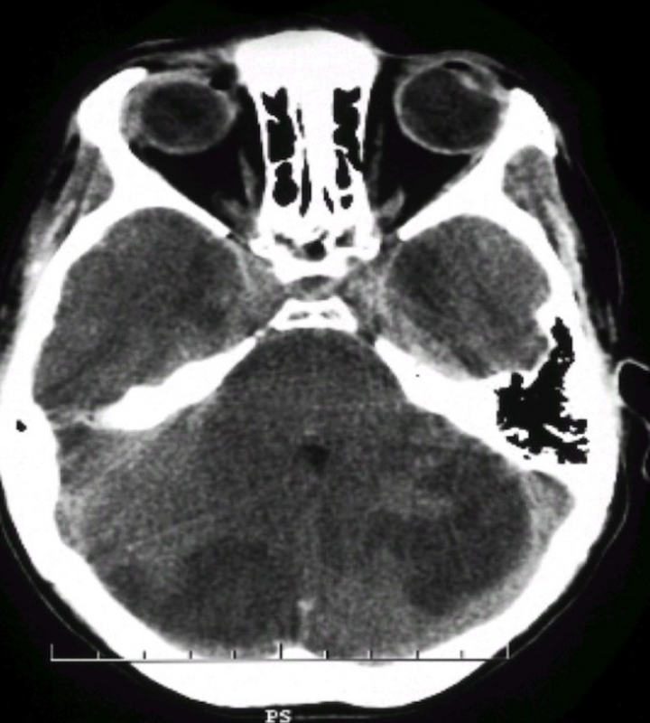
If you go back to the CT scan this is exactly what can be seen. There are bilateral areas of low attenuation in the cerebellum which are certainly the cause of this man's symptoms.
The causes include malignancy (primary, secondary or haematologic), multiple posterior circulation CVAs, cerebral vasculitis and CNS infection (although in the absence of other symptoms we would suggest this is quite unlikely).
You're probably sick of us making the same point every week - the written information provided in the examination question is every bit as important as the xray, blood gas or CT scan that accompanies it. Put together, they form a complete clinical picture that you should be able to interpret. In fact, as we have already discussed the prop is probably the lesser of the two components in terms of importance. In this case, it's very difficult to interpret without looking at the clinical information.
Don't forget: listen to what the examiners are telling you!
An otherwise asymptomatic 45 year old with vertigo and ataxia is code for:
- a cerebellar lesion
- a brain tumour, or other lesion that's probably not a bleed.
If you go back to the CT scan this is exactly what can be seen. There are bilateral areas of low attenuation in the cerebellum which are certainly the cause of this man's symptoms.
The causes include malignancy (primary, secondary or haematologic), multiple posterior circulation CVAs, cerebral vasculitis and CNS infection (although in the absence of other symptoms we would suggest this is quite unlikely).
You're probably sick of us making the same point every week - the written information provided in the examination question is every bit as important as the xray, blood gas or CT scan that accompanies it. Put together, they form a complete clinical picture that you should be able to interpret. In fact, as we have already discussed the prop is probably the lesser of the two components in terms of importance. In this case, it's very difficult to interpret without looking at the clinical information.
Don't forget: listen to what the examiners are telling you!
For the record the examiner's comments in the report were:
- The overall pass rate for this question was 48 / 82 (58.5%).
- A CT scan is performed. A CT scan showing bilateral areas of decreased attenuation in the cerebellum is shown.
- This proved to be a testing question that highlighted many candidates’ inability to interpret CT scans.
- It was considered to be an excellent test of consultant level knowledge.
- To pass candidates needed to accurately report the areas of decreased attenuation and relate this to possible causes including cardiac, embolic, vascular and malignancy. Appreciating possible causes would then lead to common sense investigations as would occur “on the floor” in the ED.
- Failures resulted from neglecting many of these issues or due to misreading the CT as a haemorrhage.